 9 weeks pregnant: Symptoms, hormones, and baby development
There are no ultrasound picture of your baby-to-be for weeks 1 and 2. While your health care providers a matter of two weeks of this towards your due date, you are not actually pregnant. Confused? You are calculated using the first day of your last menstrual period. Obviously you are not pregnant at the time, but it's the best reference you have any health care provider to estimate the arrival of a baby (until you get an ultrasound, which can provide the maturity date is more accurate).
fetal size: No measurable
Milestones fetal development: Fertilization
What You're Seeing: This week is. At some point, the sperm joins with the egg because it makes its way from the ovary through the fallopian tube and then into the uterus. Fertilization occurs in the fallopian tube. Once together, the cells begin to divide rapidly so that next week, the sonogram may be able to catch the baby-to-be early during ultrasound
The size of the fetus :. Unmeasurable
Milestones fetal development:
What You Seeing were: The small circle in the middle of the sonogram may not seem like much, but it was a little bag is a kind of baby cocoon called gestational sac. The cells that make up this bag will begin to specialize. Some cells will be a part of. Some will form the amniotic sac will fill with fluid to protect your developing baby. other cells destined to form everything from eyelashes gently to the muscle and skin. But it is still far
The size of the fetus :. 1/18 1/16 inches (about the size of a pen dot)
fetal Development Milestones: Cells that will form the heart and central nervous system are developing
What You're Seeing. dark area is liquid filled sac. Finally, it will be replaced by a fluid sac that contains the baby-to-be will live in for the next few months you. White circle in a liquid called the yolk sac. Before the placenta is fully formed, the yolk sac role in providing all the nutrients a baby-to-be needs to grow. Adjacent to the egg yolks, a little + sign indicates a very early embryo. Sonogram measures the length of the embryo (crown-rump length or CRL) to confirm or revise the estimated due date of your LMP, or to evaluate the growth of the embryo
The size of the fetus :. 1/6 to 1/4 inch
fetal Development Milestones: baby-to-be take tucked C-shape. Head, legs, and cord shape. Blood pumping through the heart
What You're Seeing. In this case, you can see a big change since the previous week's first trimester. Baby-to-be curve inward, with the umbilical cord in the middle. head appears on the right side of the image. small shoots can be seen where the arms and legs will eventually develop
The size of the fetus :. 1/6 to 1/4 inch
Milestones fetal development: There is a measured heart rate.
that you Saw: Here, the sonographer showed. The upper part shows the placement of the image of the gauges on the ultrasound machine called M-mode through pictures heartbeats. This movement of the tool shows from time to time, which is displayed at the bottom of the image. , The figure below shows how the baby's heart rate is calculated
fetal Size: 1/4 to 1/3 inch long, or about the length of your pinkie fingernail
The development of the fetus Milestones: The head grows bigger and structures that will shape the brain can be identified. Nostrils and lens of the eye develops
What You're Seeing. During this week of the first trimester, you can see the baby-to-be developed in the bubble in the gestational sac. Bubble around the embryo is amniotic fluid-filled amniotic cavity. This liquid environment gives room your baby grow and develop and move. Amniotic fluid also cushions your baby-to-be of the external pressure on the stomach. Black areas on the head is part of the neural tube develop
fetal Size: Length, 0.6 inches ;. weight, 0.04 oz
Milestones fetal development: developing baby's hands and feet. Fingers begin to form, but still joined together. Elbows and ears begin to form. Baby-to-be's body, arms and legs are getting longer. Small, jerky movements (seen on sonograms)
What You're Seeing. In this figure, the embryo is lying on his back with his head on the right side of the screen. In the now familiar C-shape, you can see that the baby-to-be head became larger during this part of the first trimester to accommodate. His brain is divided into three main parts: the forebrain, midbrain and hindbrain. As in previous weeks, the brain behind can be seen as dark areas on the back of the head of the embryo
The size of the fetus :. Long, almost 3/4 inches; weight, 2 g
fetal Development Milestones: facial features such as eyelids and ears continue to develop
What You're Seeing. The embryo appears at the bottom left of the picture with your head. The arms and legs are not visible from this angle, but the umbilical cord can be seen extending from the baby's stomach on the way to the placenta. sonogram has marked this embryo, which will help to confirm or revise the estimated due date of LMP. amniotic fluid (the dark area) surrounds the developing baby
The size of the fetus :. Long, about 3/4 of an inch; weight, 2 g
The development of the fetus Milestones: big baby's forehead, and chin backward. baby's toes are fused together
What You're Seeing. This picture gives you a sneak peek at the interaction between mother and baby during. embryo lying on his back with his head on the right side. His heart is a blue area. Stretching the umbilical cord of the baby's abdomen develops into the placenta, and red and blue colors in the cable is the blood going to and from the placenta, where it picks up oxygen and nutrients
The size of the fetus :. Long, 1.22 inches; weight, 0.14 oz
Milestones fetal development: developing eyelids, ears and the baby is fully formed (but not yet in a position). neck shape. Fingers and toes become more defined
What you see :. Would you look at this picture that the baby-to-be you are looking for more and more. arms and legs visible and recognizable profile can be seen. Bright white area in which the facial bones profile
The size of the fetus :. Long, 1.22 inches; weight, 0.14 oz
The development of the fetus Milestones :. Rudimentary forms of all organs are present, and the cartilage begins to harden and turn into bone. At the end of this week, your embryo becomes a fetus
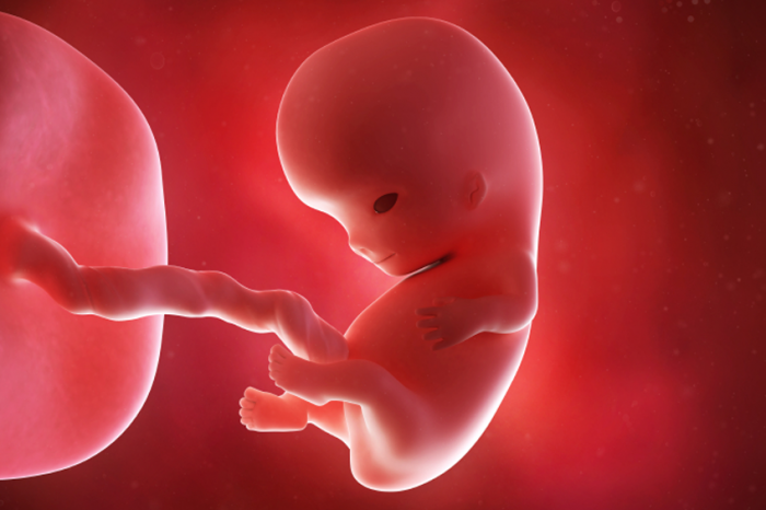
What You're Looking at :. This 3D image of your developing baby show how lifelike she appeared at this early age. Note that the baby-to-be tucked into a c-shape, with his head towards her stomach and arms and legs jutting out. , It looks away from the baby's abdomen to the placenta
fetal Size: Length, 1.61 inches; weight, 0.25 oz
fetal Development Milestones: Chin and neck evolved. become more defined. baby's ears move higher in the head
What You're Looking at :. Baby-to-be lying on his back with his head on the left side of the image and legs pointing up. From this figure, you can see that the neck grew, his big head separates from the rest of the body. His head still make up more than 50% of the size of his body, which is normal. facial bones were again seen as a bright white area in the profile
The size of the fetus :. Long, 1.61 inches; weight, 0.25 oz
The development of the fetus Milestones: The fingers and toes now. which form the baby's genitals but is not visible by ultrasound
What You're Seeing. In this 3D image, notice that the baby's delicate facial features are more visible. Muscles and bones are building in the arms and legs baby. baby umbilical cord has been slung over one shoulder. A close look also reveals tiny fingers and toes. If the image you live, you will be able to see the jerky movements developing baby is
The size of the fetus :. Long, 2:13 inch crown rump; weight, 0.49 oz
fetal Development Milestones: Fingernails and toenails begin to form. Genitals continue to develop well, though not visible on ultrasound. Kidney started functioning. And the baby may
What You're Seeing: With a baby in profile and head on the right side, you can see that the profile of the face became more and more like what you would expect to see in a newborn. Development of the baby had one hand in front of her face as if to shield his eyes
The size of the fetus :. Long, nearly 3 inches crown to rump; weight, nearly 1 ounce
Milestones fetal development: Kidney and urinary tract function. baby's fingerprints have been formed and he continued to suck his thumb. , Teeth buds are developing
What you see: In this profile shot, notice that the baby-to-be lying with his bottom on the left side of the picture and head to the right. (Although the fetus is referred to as "he" here, a sonogram may or may not be at this point.) His legs clearly visible raised, knees bent. Line in the middle of the profile is the measurement of this sonogram baby's crown-rump length (CRL). With this measurement, sonograms were able to determine the age of your baby.
All images on this slideshow are provided by sonographers from Johns Hopkins Maternal-Fetal Diagnosis and Treatment Center. We thank Christine Bird, BS, RDMS, RVT, head sonogram abortion, and Jude Crino, MD, medical director, for their assistance with this project.
For example ultrasound and more information about your baby, be sure to visit and.
Done with your first trimester? Click here for additional prenatal ultrasound:
For more information about the ultrasound and, check out the following resources:
Parents can receive compensation when you click and buy from a link contained in this site.
 You are 9 Weeks and 1 Day Pregnant - FamilyEducation
 9 Weeks Pregnant: Symptoms, Belly Pictures & More | BabyCenter
 9 Weeks Pregnant Symptoms, Ultrasound, Belly, & More - Your Baby ...
 9 Weeks Pregnant | Your Baby & You During Week 9 of Pregnancy ...
 Pregnancy week 9 | Parent24
Posting Komentar
Posting Komentar