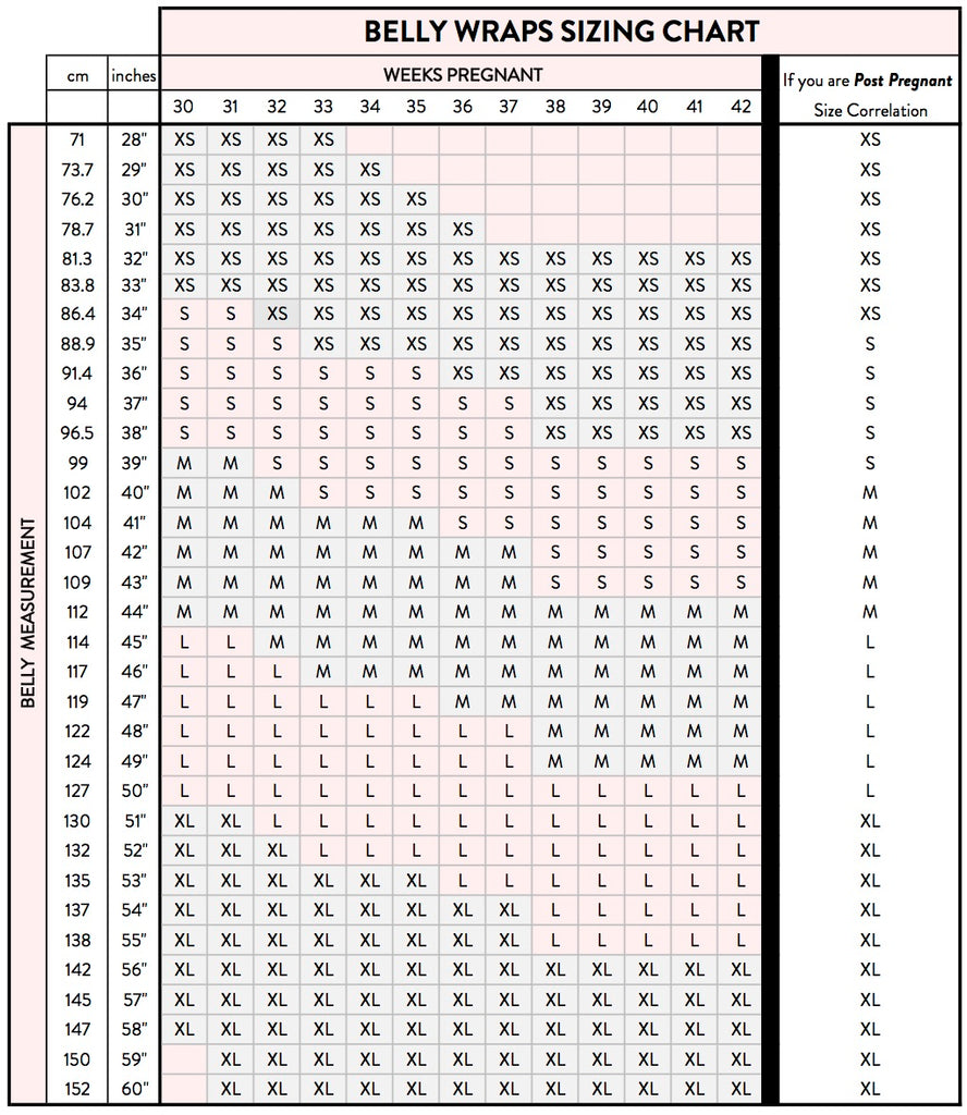
 belly measurements chart - Katan.vtngcf.org
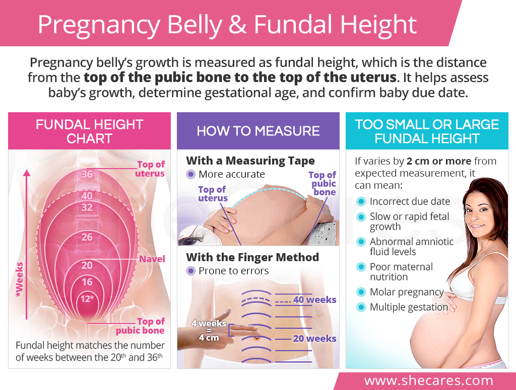
In this study session, you will learn how to perform key measurements must be performed at each antenatal visit - measuring the height of the peak of the mother's womb as a way to assess whether the baby is growing normally. We teach two ways of doing this - using your fingers, and use a soft measuring tape. This will allow you to estimate the pregnancy stage she has reached, and check the accuracy of the due date is calculated from the mother's last normal menstrual period. Then we discuss the possible reasons for the uterus to grow too fast or too slow, and what actions you should take if you suspect something might be wrong
When you have studied this session, you should be able .:
10.1 Define and use correctly all of the key words printed in bold. (SAQ 10.1)
10.2 Know how to measure the height of the fundus using gentle finger and a measuring tape. (SAQ 10.1)
10.3 Interpreting fundus measurement to assess normal fetal growth in relation to gestational age. (SAQ 10.2)
10.4 Identify possible causes normal fundus height measurements and take appropriate action. (SAQ 10.3)
The purpose of measuring the height of the mother's womb is to determine whether the baby is growing normally at any stage of pregnancy. When you measure the womb, you check to see where the top of the uterus is.
signs Healthy
signs Warning
Do you remember what the dome area at the top of the uterus is called? (You learned this in Study Session 3.)
This is called the fundus.
When you are measuring how high the uterus has reached a peak in the mother's abdomen, you measure the height of the fundus. This is a much more accurate estimate of the weight of maternal fetal growth. Measuring fundus will show three things:
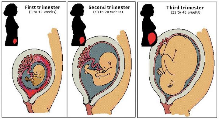
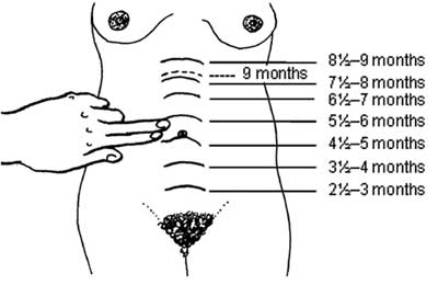
As the baby grows in the womb, you can feel the uterus grows larger in the mother's abdomen. The upper part of the uterus moves around two fingers or 4 cm higher each month (Box 10.1).
In about three months (13-14 weeks), the upper part of the uterus is usually just above the mother's pubic bone (where her pubic hair begins).
At about five months (20-22 weeks), the upper part of the uterus is usually right on the mother navel (umbilicus or navel).
At about eight to nine months (36-40 weeks), the upper part of the uterus almost to the bottom of the rib mother.
Babies may drop lower in the weeks before the birth. You can look back at Figure 7.1 in Study Session 7 to see a diagram of the fundus at various weeks of pregnancy.
For a taste of the uterus, have the mother lie on her back with some support under her head and knee. Explain to him what you will do (and why) before you start touching her stomach. You touch should be firm but gentle. Walk your fingers to the side of the stomach (Figure 10.1) until you feel under the skin on her stomach. It will feel like a hard ball. You can feel the top by curving your fingers gently into the abdomen.
If the top of the uterus is below the navel, measure how many fingers below the navel. If the top of the womb is above the bellybutton, measure how many fingers above the navel.
Look carefully at Figure 10.2. If the baby is growing normally, with how many fingers-width should increase in the uterus in the second trimester (3-6 months of pregnancy, or 15-27 full week)?
High fundus should increase by 6 finger-width (two fingers in every month) in the second trimester.
How many fingers above the navel should be top of the uterus is located at 7 months' gestation?
See Figure 10.3 for the answer.
How do you explain the position of the dotted line at 9 months in Figure 10.2, which is below the line indicate the height of the fundus in 8 ½ to 9 months?
Babies may drop lower in the weeks before birth (see back at Box 10.1).
Look at the diagram in Figure 10.4 (a) and (b). How many weeks pregnant women in each case, based on the method of measuring finger fundus is shown in Figure 10.2?
In Figure 10.4 (a) woman is about 4 ½ months pregnant. In Figure 10.4 (b) he is about 6 ½ months pregnant (three fingers above the navel).
When you measure the height of the fundus at each antenatal visit, write down the number of fingers used to measure the height of the uterus in women antenatal record card, Put the '+' (plus) sign in front of the number if the upper part of the uterus above the navel. Put a '-' (minus) sign in front of the number if the upper part of the uterus is below the navel
How do you record the measurements are shown in Figure 10.4 (a) and (b).?
measurements in Figure 10.4 (a) shall be recorded as -2. Measurements in Figure 10.4 (b) will be +3.
You need to realize that the finger method for estimating gestational age (number of weeks / months of pregnancy) has some limitations that affect accuracy.
Look at your own hand. Can you suggest why the finger method may give different estimates of pregnancy if two different health workers use this method to measure the height of fundus this same woman?
Because of large variations in the thickness of our fingers, there can be up to three weeks fundus differences between measurements of the same woman made by two different people. (This is known as 'variation between observers', ie the variation between different observers.)
Even if the actions of health workers at the fundus of the same woman several times on the same day, the answer may be different each time, because the method is not very precise finger. (This is known as 'variations of intra-observer', ie variation by a single observer at different times.)
Finally, you may have noticed that the distance between the symphysis pubis (pubic bone) and the umbilicus (navel) varies among women when they are pregnant, and these variations affect measurement accuracy fundus using fingers. For example, it is assumed that the distance between the symphysis pubis and the umbilicus is 20 cm at 20 weeks gestation, but can be as long as 30 cm and as short as 14 cm.
To overcome this limitation, it is recommended that you measure the height of the fundus using a soft measuring tape if you have one, as described next.
You can use this method when the top of the uterus grow as tall as the woman's navel.
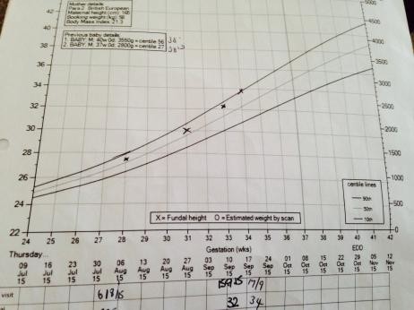
During the second half of pregnancy, the uterus size in centimeters closer to the number of weeks that the woman was pregnant. For example, if it has been 24 weeks since the last normal menstrual period, the uterus will usually measure 22-26 cm. Rahim should grow about 1 cm every week, or 4 cm every month.
Doctors, nurses and midwives are taught to count pregnancy by weeks instead of months. They start counting on the first day of the last normal menstrual period (LNMP), even though she was probably pregnant two weeks later. Counting this way makes the most of the long gestation of 40 weeks (or you can say a normal pregnancy is 40 weeks).
If you are measuring correctly and you do not find the top of the uterus where you expect it to be, based on the date the lady gave you for LNMP her, it could mean three different things:
There are several reasons why the maturity of thought of LNMP could be wrong. Sometimes women do not remember the date of their LNMP properly. Sometimes a woman misses her menstrual period for other reasons, and then gets pregnant later. This woman really could be less pregnant than you think, so that the uterus is smaller than you expected. Or sometimes a woman has a little bleeding after she was pregnant. If he considers that it LNMP, this woman will be one or two months more pregnant than you thought. the uterus will be greater than you expect.
Remember the due date is not appropriate. Women often give birth to 2 or 3 weeks before or after their due date. It is usually safe.
If the due date does not match the size of the uterus during the first visit, take notes. Wait and measure the womb again in two to four weeks. If the uterus grows about two finger-width or 1 cm per month, the maturity date of the feeling you get on the uterus may be true. The due date you got by counting from LNMP probably one
If the uterus grows more than 2 fingers in a month, or more than 1 cm in the week, several different possible causes :.
If you think there might be twins, even if you can find only one heartbeat, refer the woman to the nearest health center.
This can be very difficult to know for sure that a mother pregnant with twins. Signs of twins is that:
We'll show you how to listen to the heartbeat of the fetus through the mother's abdomen in Study Session 11. For the moment, we are focusing on a twin as pOssible reasons for the uterus to be larger than expected. Here are two ways to try to hear the heartbeats of twins:
Since the birth of twins is often more difficult or dangerous than single births, safer for women to go to the hospital to give birth. Since twins are more likely to be born early, she should try to have transportation ready at any time after the 6th month. If the hospital away, the mother may want to move closer in the last months of pregnancy. Be sure to have a plan for how to get help in an emergency.
You learn about the warning signs of diabetes in Study Session 9.
If a woman has all the warning signs of diabetes, what do you expect to find?
Refer the woman to a health center if you suspect she may have diabetes.
He suffered from gestational diabetes in the past. One of her past babies born very large (more than 4 kilograms), or sick or died at birth, and no one knows why. He's fat. He was thirsty all the time. He has often itching and the smell coming from her vagina. the wound healed slowly. He had to urinate more frequently than other pregnant women. uterus is larger than normal for how many months she has been pregnant. He has sugar in the urine when you do a dipstick test (Section 9.8.1 of the Study Session 9).
Too much water (amniotic fluid) is not necessarily a problem, but can cause the uterus to stretch too much. Then the uterus can not contract enough to push the baby out, or to stop bleeding after childbirth. In rare cases, it may mean that the baby will have a birth defect. Try to refer the woman to the nearest health facility that could give him a sonogram (ultrasound) if the uterus is too big and you measure the twins are not suspects.
Sometimes a pregnant woman, but the tumor grew not a baby. It is called a molar pregnancy (Figure 10.7). Spotting blood and tissue (sometimes shaped like grapes) can be removed from the vagina.
If you detect signs and symptoms of a molar pregnancy, refer her to the hospital as soon as possible. Tumors can be cancerous and kill it, sometimes very quickly. A surgeon can remove the tumor to save the lives of women
Other signs of a molar pregnancy is that :.
The slower growth can be a sign of one of these problems:
If you do not have the proper equipment to check blood pressure, and the uterus grows too slowly, referring him to the nearest health center for evaluation.
If you suspect that the baby may have died, refer the mother to a health center for the stillbirth.
If a pregnant woman five months or so, ask if he had felt the baby move recently. If the baby does not move for two days, something may be wrong. If the mother is more than seven months pregnant, or if you hear the baby's heartbeat on a previous visit, listen to the heartbeat again.
If the woman reported no fetal movement and you can not hear the heartbeat, the baby may have died. If so, it is important for the baby's death (stillbirth) to be delivered immediately, because a woman may bleed more than other mothers, and she is at greater risk of infection.
When a mother loses a baby, she needs love, care and understanding (Figure 10.8). Make sure that he does not go through labor alone. If she gave birth to a baby dies in the hospital, someone who he believed had to stay there with her during the birth.
In this study session, you have learned how to measure the height of the fundus, using your fingers and the tape measure. You also have to learn to interpret your measurements and take appropriate action. In the next study session you will learn how to assess the baby's position by palpating (feeling) the mother's abdomen and listen to the fetal heart rate position.
In Study Session 10, you have learned that:
Now that you have completed the study session, you can assess how well you have achieved learning outcomes by answering the questions below Case Study 10.1. Write your answers in your Study Diary and discuss them with your Tutor at the next Study Support Meeting. You can check your answers with the Notes on Self-Assessment Questions at the end of this module.
Abebech is a pregnant woman, the duration of the pregnancy based on her last normal menstrual period (LNMP) is six months. When you examine him, you can feel that the fundus is his four fingers above the navel and you can hear the fetal heartbeat clearly.
Is Abebech baby's gestational age based on height measurements of the fundus is consistent with gestational age calculated from his LNMP?
gestational age based on fundus is one month more than expected from LNMP date. Therefore, the uterus is larger than expected from LNMP date.
What possible explanation can you give for your findings in this Abebech case, and what actions should you take?
The uterus may be larger than expected due date may be wrong LNMP, and Abebech actually seven months pregnant. This is not a problem, but it is important to investigate other possible explanations. For example, he may have too much amniotic fluid (water) that surrounds the baby in the womb; You should refer to a medical facility where he can have an ultrasound to find out if this is the case. Or he could have a twin pregnancy. You can hear the fetal heartbeat clear, so get someone else to help you listen Abebech stomach to see if you can hear two fetal heartbeats. If you suspect he's having twins, referring him to the nearest health facility.
Except for third party materials and / or otherwise stated (see) content in OpenLearn released for use under the provisions. In short it allows you to use content worldwide without payment for non-commercial purposes in accordance with a non-commercial Creative Commons Share Alike license. Please read this license in full together with the term OpenLearn and conditions before making use of the content.
When using your content must attribute our (Open University) (OU) and every author is identified in accordance with the requirements of the Creative Commons License.
Part Acknowledgments used to list, among others, third party (Proprietary), licensed content that is not subject to a Creative Commons license. Right of ownership The content should be used (maintained) intact and in context with the contents at any time. That Acknowledgments section is also used to bring your attention to any other restricted special that may apply to the content. For example, there may be times when a Creative Commons Non-Commercial ShareAlike license is not valid for any of the content even if it is owned by us (which OU). In this manner, unless otherwise stated, the content can be used for personal and non-commercial use. We also have been identified as other materials Proprietary included in a content subject Creative Commons License. It is: OU logo, trade name and may extend to certain photographic and video images and sound recordings and other materials that may be brought to your attention.
Unauthorized use of any content may be a violation of the terms and conditions and / or intellectual property laws.
We reserve the right to change, alter or bring to an end the terms and conditions provided here without notice.
All rights fall outside the terms of the Creative Commons licenses is maintained or controlled by the Open University.
Head of Intellectual Property, The Open University
 stomach measurement chart - Katan.vtngcf.org
 belly measurements chart - Katan.vtngcf.org
 Supportive Belly Band | Maternity & Nursing Wear | Medela | Medela
 Maternity Size Chart | Motherhood Closet - Maternity Consignment
 Pin on baby shower mickey
 Sizeinfo Maternity
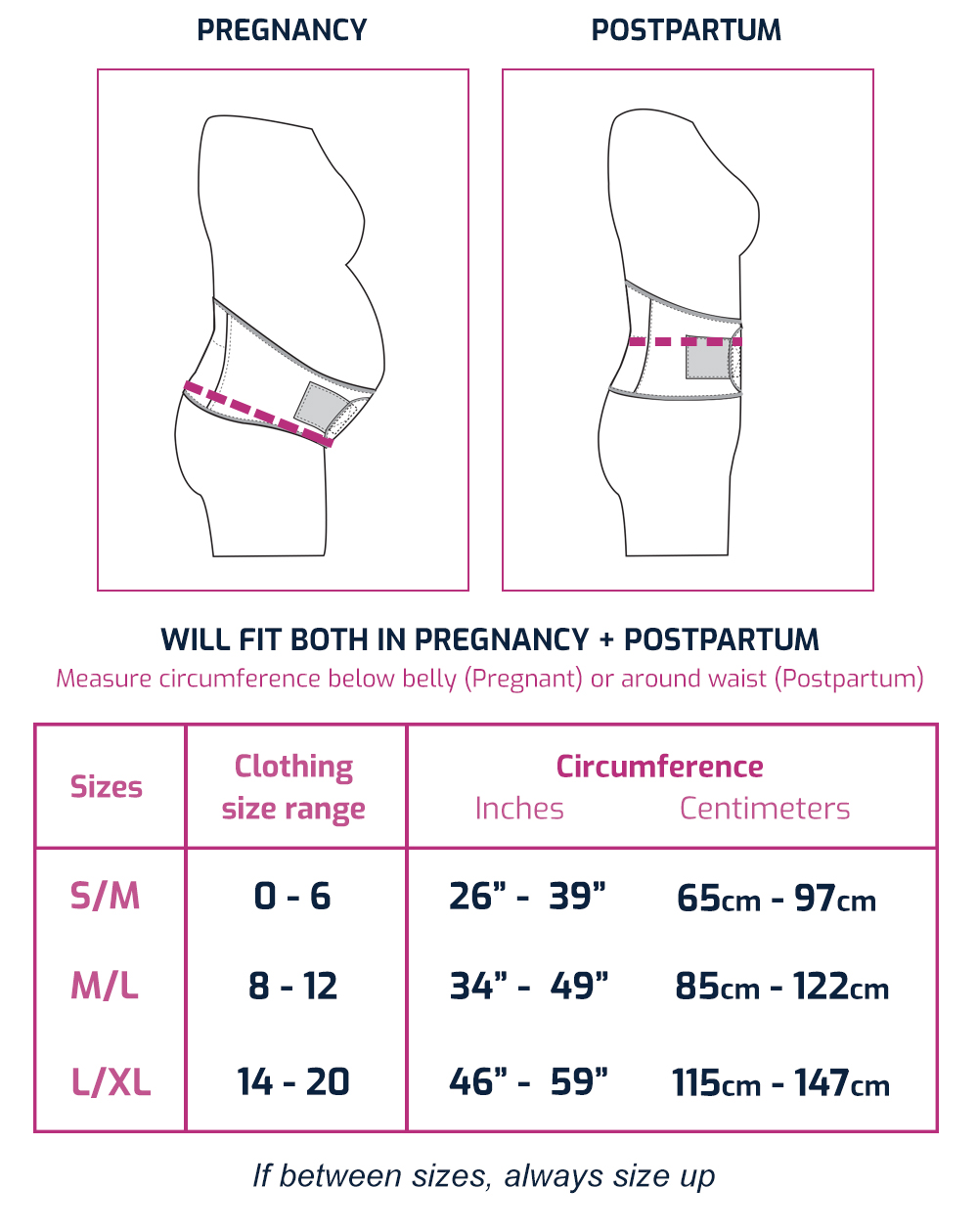 Pregnant Belly Size Chart - Blackmores Pregnancy
 stomach size during pregnancy chart - Catan.vtngcf.org
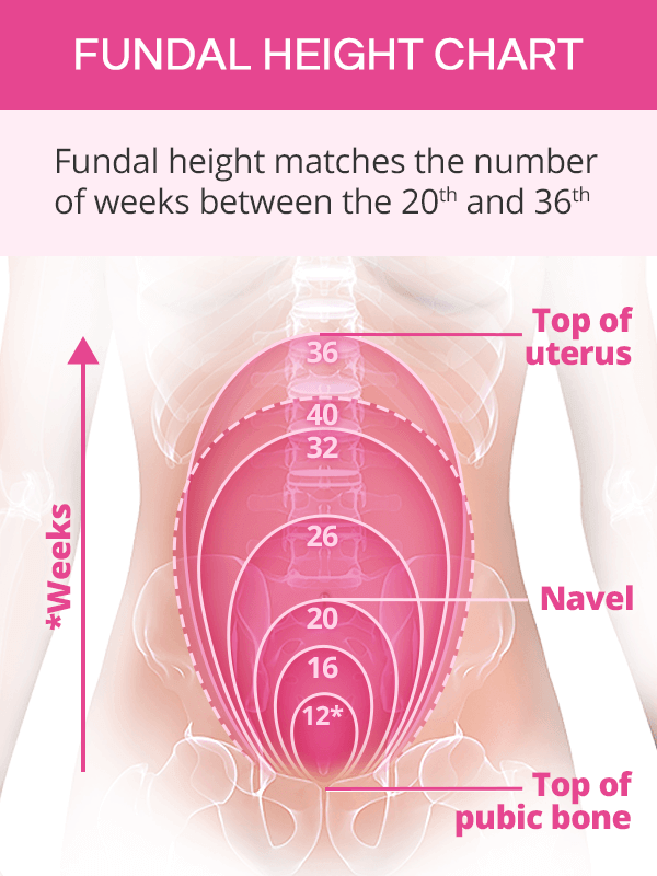 Pregnancy Belly and Fundal Height | SheCares
 Pregnancy Belly Measurements Chart - Unborn baby growth chart yok ...
 Belly Size during Pregnancy - Week by Week Chart
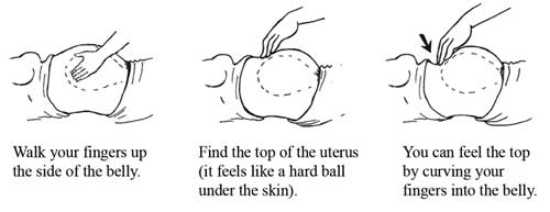 Antenatal Care Module: 10. Estimating Gestational Age from Fundal ...
 Pin op Maternity/Pregnancy
 Pregnant Measurement – Fashion dresses
 Pregnant Belly Size Chart And Shape: Things You Should Know
 Pregnant Belly: Does Size Matter?
 Pregnant Belly Measurements - Blackmores Pregnancy
 pregnancy stomach measurement chart - Matan.vtngcf.org
 Pregnancy Belly and Fundal Height | SheCares
 Head Circumference Calculator | babyMed.com
 Pregnancy by week photos
 How to Measure Fundal Height: 15 Steps (with Pictures) - wikiHow
 Belly Bandit- Women's Maternity Belly Boost — Cute Little Me!
 Fetal Growth Examples
 Uterus During Pregnancy: Its Size, Changes And Role
 Estimation of Fetal Weight
 Pregnant Belly Measurements Chart - Blackmores Pregnancy
 Belly Size during Pregnancy - Week by Week Chart
 Maternity Top Short Sleeve Funny Pregnancy Tee Cute Pregnant Women ...
 12 Best Pregnancy Tracker Apps 2020 - Baby Apps
_046ab245-f10c-47fe-808e-9e5cb12b15a1-9ce4d7.jpg) Having a small baby | Pregnancy Birth and Baby
Having a small baby | Pregnancy Birth and Baby Pin on Pregnancy/Childbirthing
 Ultrasound measurement of fetal kidney length in normal pregnancy ...
 Pregnant Belly Measurement - Blackmores Pregnancy
 Pregnancy Size by Week | LoveToKnow
 Fetal Growth Examples
 Baby Belly Band Original Pregnancy and Post Partum Belt for ...
 Ultrasound measurement of fetal kidney length in normal pregnancy ...
 UpSpring BumpTube Pregnancy Belly Band for Pregnancy Support at ...
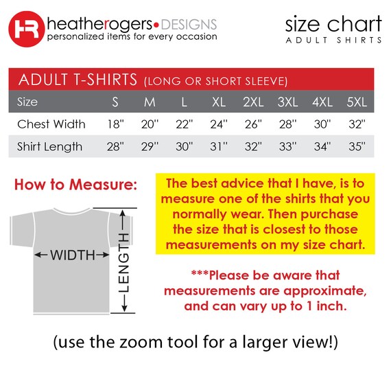 The Man Behind the Belly Humor Dad Shirt / Dad Pregnancy | Etsy
 Dinámica de Crecimiento del Estómago en el Período Fetal Humano ...
 Leonisa High Waist Post-Pregnancy Adjustable Panty Belly Wrap Canada
 Antenatal Care Module: 10. Estimating Gestational Age from Fundal ...
 How to Measure Fundal Height: 15 Steps (with Pictures) - wikiHow
 25 Weeks Pregnant: Symptoms, Tips and Fetal Development | Pampers
 Confused by tummy measurements and Centile graph ? | Netmums
Posting Komentar
Posting Komentar